


Check out this blog post by Parkland College, which talks about some of the new technology they’re utilizing. Seriously awesome.
They’ll be on site for the Pygmalion Festival‘s TECH Demo Day on Friday, September 25th at Krannert Center.
Below, Biology Professor Lori Garrett shares how Parkland’s new Anatomage table, with its high-tech virtual dissection technology, is helping students learn. Plus, check out an exerpt from her upcoming video to be shown during the Pygmalion Tech Fest.
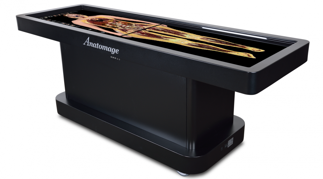
**Parkland is a presenting partner of the Pygmalion Festival, September 23-27, which includes a Tech Festival on Friday, Sept., 25 at Krannert Center in Urbana. The Tech Festival is FREE for all Parkland students with a valid ID.**Parkland is amazingly fortunate to have an Anatomage digital dissecton table. These state-of-the-art, high tech tables were developed primarily for the medical field, and there are only about 500 in use worldwide, with only a little over 200 currently in use in the U.S. Those are primarily located in hospitals and medical schools. It’s such a high-tech piece of computerized equipment that I attended a two-day User Group meeting in San Francisco in August for in-depth training, and we’re just starting to really appreciate all we can do with it ourselves.
What the Table Does and What We Can Do With It
The Anatomage is like two giant, touch-screen computer monitors with highly sophisticated software behind them. The image banks were developed at Stanford University and are based off of real human CT scans and anatomical models. It provides us with life-size 3D renderings of three different individuals, and we can dissect through them. We can approach the anatomy from the surface and scroll down through the tissue layers, or isolate individual organs and organ systems. Various icons allow us to cut through, or section, any of the body parts, view X-ray images, isolate organ systems, see soft tissues, and more—and everything’s rendered in three dimensions, rotatable, and zoomable. We can add labels, place pins on structures for examinations, and add our own notes all on screen.
We’re really excited for the promise the software holds for advancing our science and medical instruction. With the Anatomage’s InVivo software program, we can take CT or MRI scans from anyone, anonymize them, and then have them digitized and rendered in 3D. This will let us use real-life case studies in a cross-curricular manner for our students moving into the health professions. We can also use the software to isolate any organs, save the digitized data, and then use 3D printing to develop our own anatomical models.
What Students Think about the Anatomage Table
Our students love the Anatomage table because of its technology. We’re integrating the table in our anatomy classes, where we already use plastic models and human cadavers. The table allows our students to learn anatomy from life-size renderings of real cadavers, which makes their cadaver study much easier. In the cadavers, we can’t isolate whole organ systems or rebuild the body like we can on the Anatomage. Being so tech-savvy, our students embrace it and need little guidance—they are used to touchscreen computers and phones.
We sometimes give tours for high school anatomy classes and let the students try the table after a brief introduction and demonstration. Being digital natives, they take to it with no effort at all.
The Anatomage allows us to bridge the gap between simulators and real people. It lets us visualize organs, vessels, tissues, and more without worrying about torn structures or extra tissues and clutter as we see in the real cadavers. The Anatomage is life-sized like our cadavers, but without the “delightful” aroma of the chemical preservatives, and we know our students really appreciate that!
Patrick Singer
Executive Editor